What We Do
The Hoon Lab combines high resolution imaging with transgenic approaches to unravel the mechanisms that regulate synaptic connectivity during retinal circuit development and also during disease and degeneration conditions. We hope to offer novel therapeutic possibilities by correlating circuit plasticity during development and disease.
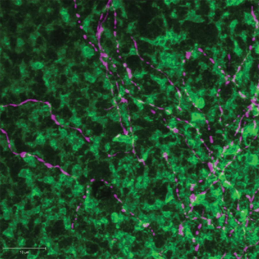
We use multi-transgenic approaches to label retinal cell types and visualize connections between retinal neurons. Shown here is a part of a GABAergic amacrine arbor (magenta) overlaid amongst bipolar cell terminals (green) that code visual information during dim-light conditions. Visualizing synaptic partners is the first step in being able to study the development, maintenance and degeneration of retinal connections.
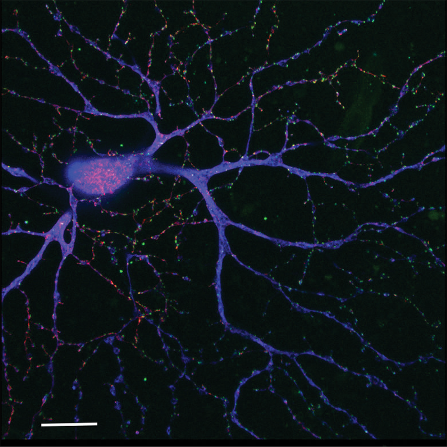
We utilize biolistic transfection techniques to label retinal ganglion cells and synapses on the dendritic arbors of these cells. Shown here is the distribution of excitatory (green) and inhibitory (red) synaptic proteins on the arbor of an ‘ON’ retinal ganglion cell that codes for increases in illumination.
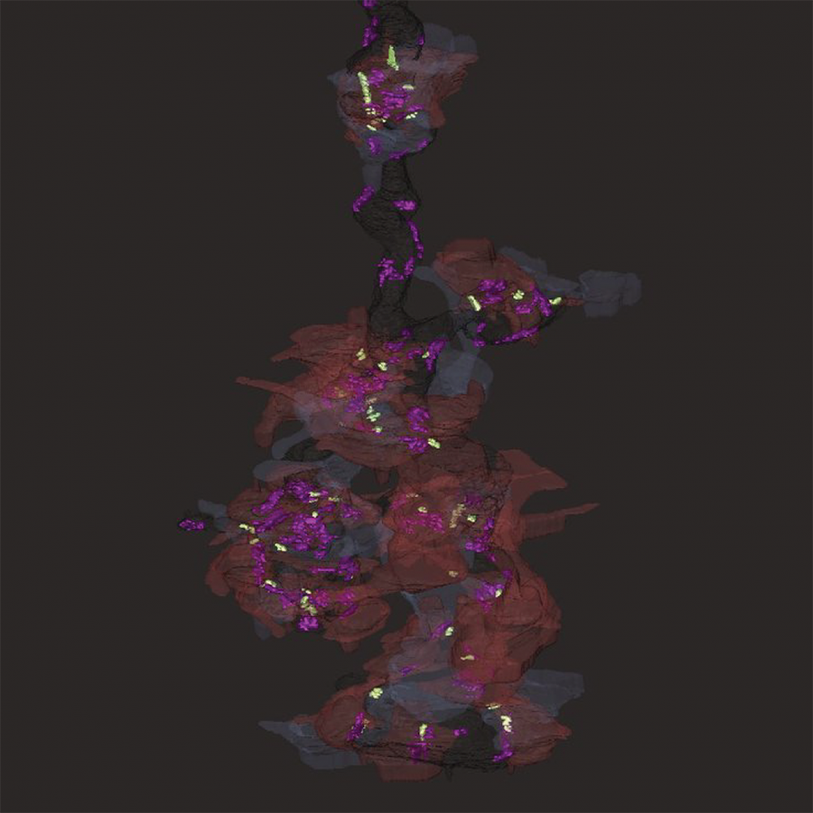
We reconstruct the ultrastructural connections between retinal neurons by electron microscopy. Shown here is a reconstructed rod bipolar cell terminal (dark grey) from mouse retina with locations of output ribbon synapses (green) and modulatory inhibitory synapses (magenta). The two amacrine partners at each rod bipolar ribbon output have also been reconstructed. The GABAergic partner is color-coded in brown and the glycinergic partner is color-coded in silver.
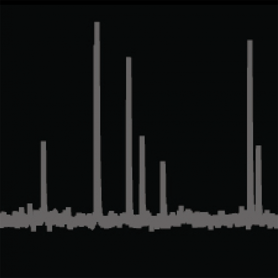
Our work correlates synapse structural organization with assessments of synaptic function. We use whole-cell patch clamp recording to assay light-driven responses from retinal neurons.